| |
| Mechanism of the development of gastric ulcer
after percutaneous
endoscopic gastrostomy |
Jiro Kanie*, Hiroyasu Akatsu**, Yusuke Suzuki****,
Hiroshi Shimokata***,
Akihisa Iguchi****
* Department of Internal
Medicine, Fukiage
Digestive Endoscopy
Center
** Department of
Internal Medicine, Sawarabi-kai
Fukushimura Hospital
*** Department of
Epidemiology National Institute
for Longevity Sciences
****Department of
Geriatric Medicine,
Nagoya
University, School
of Medicine
|
|
| Endoscopy 2002; 34(6): 480-482 |
|
|
|
| Summary |
Background and study aims: The present study was carried out in order
to elucidate the mechanism of
the development
of gastric ulcer, one of the
serious complications
of PEG tube placement.
Patients and methods: This retrospective study included 92 patients
who underwent gastric endoscopy
after PEG
tube placement. Gastric ulcers
detected at
gastroscopy were examined in
relation to
the length of the protrusion
from the intragastric
bumper of the PEG tube’s intragastric
bumper
and the use of histamine H2-blocker.
Results: Gastric ulcer was found in 9 of the 92 patients,
and in all nine the ulcer was
found on the
posterior wall of the gastric
body, where
the tip of the PEG tube was attached.
Seven
of the 21 patients (33.3%) who
had a PEG
tube with a long protrusion from
the intragastric
bumper developed gastric ulcer.
By contrast,
only two of the 71 patients (2.8%)
who had
a PEG tube with a short protrusion
developed
gastric ulcer. The use of H2-blocker
had
no significant impact on the
development
of gastric ulcer.
Conclusions: The occurrence of gastric ulcer after PEG
placement was attributable to the shape of
the PEG tube within the intragastric space,
and not to the use of H2-blockers, suggesting
that appropriate placement of the PEG tube
is an important factor in preventing gastric
ulcer.
|
| Introduction |
The value of tube feeding with percutaneous
endoscopic gastrostomy (PEG) has been clearly
recognized, and PEG tube feeding is now widely
used in elderly patients with dysphagia due
to cerebral apoplexy or senile dementia;
nasogastric tube feeding is also still widely
used [1-3]. With the widespread use of PEG
feeding, there have been reports of several
complications peculiar to PEG feeding [4-8],
as well as reports on ways of preventing
these [9-12]. However, these reports have
been limited to complications during the
acute postoperative phase, with the exception
of buried bumper syndrome in the chronic
postoperative phase [13-15]. There have been
few reports of other complications during
the chronic phase, particularly the development
of gastric ulcer as a severe complication
of PEG tube placement. The aim in the present
study were to investigate the incidence of
gastric ulcer detected at gastroscopy after
PEG placement, and to examine the contribution
of two possible factors - the shape of intragastric
bumper and the use of histamine H2-receptor
antagonists (H2-blocker).
|
| Patients and Methods |
Patients
The study included 92 patients (29 men,
63 women, mean age; 78.3, range; 39-97),
who underwent gastric endoscopy after PEG
tube placement. Gastroscopy was carried out
when the tubes were being exchanged. The
disease backgrounds for all the patients
included are shown in table 1. Gastric endoscopy
was carried out a mean of 249 days (range
6-1833 days) after PEG tube placement during
tube exchange, except in patient who presented
with clinical symptoms of gastrointestinal
bleeding. Non of the patients had any past
history of gastric ulcer, and no gastric
ulcer was detected when the initial PEG placement
was carry out. The patients or their relative
agreed to the gastroscopy examinations after
PEG tube placement, and provided written
informed consent after receiving a sufficient
explanation of the procedure.
Methods
The 92 patients were divided into two groups
on the basis of the length of tube protruding
from the intragastric bumper of the PEG tube.
Group 1 consisted of patients in whom the
tube protruded 5 mm or more from the intragastric
bumper, and group 2 included those in whom
the protruding tube was less than 5mm. Two
different types of bumper were used in both
groups (balloon bumper and Malecot bumper
in group 1 balloon bumper and silicon bumper
in group 2). The numbers of patients in the
two groups are shown in Table 1. The patients
were also divided according to their use
of H2-blockers. H2-blocker administration
was started after the onset of stroke to
prevent complications such as Cushing’s
ulcer, and was continued up to the time gastroscopy
was performed.
Statistical analyses were carried out
using
Fisher's exact test.
|
| Table 1. |
Characteristics of the Patients who underwent
Gastroscopy after PEG tube placement of a
percutaneous endoscopic gastrostomy(PEG)
tube |
|
| |
|
Group 1*
(n=21) |
Group 2**
(n=71) |
Total
(n=91) |
|
| Disease |
|
Cerebral Infarction |
6 |
31 |
37 |
| Dementia |
8 |
26 |
34 |
| Cerebral hemorrhage |
4 |
2 |
6 |
| Subarachnoid hemorrhage |
0 |
3 |
3 |
| Brain contusion |
1 |
2 |
3 |
| Brain anoxia |
0 |
2 |
2 |
| Amyotrophic lateral sclerosis |
1 |
1 |
2 |
| Parkinson's syndrome |
0 |
1 |
1 |
| Gastric cancer |
0 |
1 |
1 |
| Progressive supranuclear palsy |
0 |
1 |
1 |
| Encephalitis |
0 |
1 |
1 |
| Brain tumor |
1 |
0 |
1 |
| Sex |
|
Male |
6 |
23 |
29 |
| Female |
15 |
48 |
63 |
Age
(y;mean, range) |
79.24
( 55-97 ) |
78.04
( 39-94 ) |
78.32
( 39-97 ) |
Interval from PEG ***
(days; mean, range) |
237
( 6-1833 ) |
252
( 13-801 ) |
249
( 6-1833 ) |
|
| * |
Group I: Protrusion from Intragastric Bumper
≧ 5mm |
| ** |
Group II: Protrusion from Intragastric Bumper
< 5mm |
| *** |
Interval between the day of percutaneous endoscopic gastrostomy and that of gastroscopy |
|
| Figure 1. |
Categorization of the PEG tubes according
to the length of the protrusion from the
intragastric bumper of the PEG tube as observed
by gastroscopy |
|
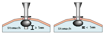 |
| a |
b |
|
|
a Group 1: protrusion from intragastric bumper
≧5mm
b Group 2: protrusion from intragastric bumper
<5mm |
|
Results |
Incidence of Gastric Ulcer after PEG Tube
Placement
Of the 92 patients who underwent gastroscopy
after PEG placement, nine (9.9%) were found
to have gastric ulcers. Among the nine patients
diagnosed with gastric ulcer at gastroscopy,
three patients in group 1 showed clinical
symptoms of gastrointestinal bleeding. The
other four patients in group 1 and two patients
in group 2 were asymptomatic. There were
no differences between the groups with regard
to complications or other confounding factors
(e.g., age, types of medication, disorders
such as respiratory, renal, or hepatic dysfunction)
capable of increasing the risk of gastric
ulcer. In all nine patients, the gastric
ulcers were located on the posterior wall
of the body of the stomach, where the tip
of the PEG tube was in contact with the mucosa.
Seven (33.3%) of the 21 patients in group
1 (long protrusion), and two (2.8%) of the
71 patients in group 2 (short protrusion)
developed gastric ulcer. The occurrence of
gastric ulcer was significantly higher in
group 1 patients compared with group 2 patients
(P < 0.05, Fisher's exact test)
|
Table 2. Relationship between PEG tube shape
and the development of gastric
ulcer
|
|
Group 1* |
Group 2** |
Total |
|
| Gastric ulcer |
7 (33.3%) |
2 (2.8%) |
9 |
| No gastric ulcer |
14 (66.7%) |
69 (97.2%) |
83 |
| Total |
21 (100%) |
71 (100%) |
92 |
|
* Group I: Protrusion from Intragastric
Bumper
≧ 5mm
**Group II: Protrusion from Intragastric Bumper <
5mm
p<0.05
; Fisher’s exact probability test
|
Effect of H2-Blocker Administration
An H2lblocker was administered
to four
of the 92 patients who underwent
gastroscopy
after PEG tube placement. Among
the 21 patients
in group 1, gastric ulcer was
observed in
one of the two patients who were
receiving
an H2-blocker, and in six of
the 19 patients
who were not receiving an H2-blocker.
In
group 2, none of the patients
who were on
an H2-blocker developed gastric
ulcer, while
two of the 69 patients who were
not on an
H2-blocker developed gastric
ulcer. The use
of H2-blockers had no significant
impact
on the onset of gastric ulcer
in either group.
|
Table 3. H2 blocker medication and stomach
ulcer outbreak risk among the
patients in
Group I and Group II
|
|
Group 1 |
Group 2 |
|
H2-blockers |
No H2-blockers |
Total |
H2-blockers |
No H2-blockers |
Total |
|
| Gastric ulcer |
1 |
6 |
7 |
0 |
2 |
2 |
| No gastric ulcer |
1 |
13 |
14 |
2 |
67 |
69 |
| Total |
2 |
19 |
21 |
2 |
69 |
71 |
|
n.s. ; Fisher’s
exact probability test
|
| Discussion |
PEG was first described by Gauderer et
al. in 1980 (16), and PEG tube placement
is highly regarded as a useful method for
managing patients who require long-term transtubu1ar
feeding. We previously reported (17) that
complications are more frequent after PEG
than reported by Jain et al. (18). In our
experience in 441 patients who underwent
PEG, there were 144 incidents of post-PEG
complications, including gastric ulcer.
Some speculations have been published regarding
the mechanism underlying the development
of gastric ulcer after PEG tube placement.
Several reports (19,20) have suggested the
possibility that contact between a nasogastric
feeding tube and the gastric wall may be
a cause of gastric ulcer. However, this mechanism
has not previously been demonstrated for
the onset of gastric ulcer in patients undergoing
PEG placement. In the present study' in all
nine patients who developed gastric ulcer
after PEG tube placement, the gastric ulcer
was observed on the posterior wall of the
gastric body, where the tip of the PEG tube
came into contact with the mucosa. None of
the 92 patients in the present study had
any previous history of gastric ulcer. In
addition, it was confirmed that the gastroscope
was aseptic for Helicobacter pylori before
the gastroscopy procedure in each patient.
It is therefore likely that mechanical stimulation
by the PEG tube on the mucosa of the stomach
led to the development of the gastric ulcers,
and this view is supported by the finding
that gastric ulcer occurred in a significantly
higher percentage of group 1 patients, in
whom the PEG tube was more likely to cause
injury to the gastric mucosa due to the longer
protrusion from the bumper.
Only four of the 92 patients studied had
received H2-blocker treatment before PEG
tube placement. However, H2-blocker administration
did not significantly reduce the incidence
of gastric ulcer. As detailed above, we would
speculate from these results that the development
of gastric ulcer after PEG tube placement
may be due to mechanical injury caused by
the PEG tube to the gastric mucosa, and that
the administration of H2-blockers may not
prevent the development of gastric ulcer.
|
| Conclusion |
Use of a PEG tube with a long protruding
tip was associated with a significantly higher
frequency of post-PEG gastric ulcer due to
contact injury to the gastric mucosa caused
by the tip of the tube. Choosing the appropriate
PEG tube may be crucial in preventing gastric
ulcer alter PEG placement.
|
| References |
| (1) |
Norton B, Homer-Ward M, Donnelly MT, et al.
A randomised prospective comparison of percutaneous
endoscopic gastrostomy and nasogastric tube
feeding after acute dysphagic stroke. BMJ
1996; 312: 13-16 |
| (2) |
Park RH, Allison MC, Lang J, et al. Randomised
comparison of percutaneous endoscopic gastrostomy
and nasogastric tube feeding in patients
with persisting neurological dysphagia. BMJ
1992; 304:1406-1409 |
| (3) |
Kanie J, Kono K, Yamamoto T, et al. Gastro-esophageal
reflex successfully treated with transgastrostomal
jejunal tube feeding. Jpn J Geriat 1997;
34: 60-64 |
| (4) |
Haslam N, Hughes S, Harrison RF. Peritoneal
leakage of gastric contents, a rare complication
of percutaneous endoscopic gastrostomy. J
Parenter Enteral Nutr 1996; 20: 433-434 |
| (5) |
Fox VL, Abel SD, Malas S, et al. Complications
following percutaneous endoscopic gastrostomy
and subsequent catheter replacement in children
and young adults. Gastrointest Endosc 1997;
45: 64-71 |
| (6) |
Petersen TI, Kruse A. Complications of percutaneous
endoscopic gastrostomy. Eur J Surg 1997;
653: 351-356 |
| (7) |
Patel AS, DeRidder PH, Alexander TJ, et al.
Candida cellulitis: a complication of percutaneous
endoscopic gastrostomy. Gastrointest Endosc
1989; 35: 571-572 |
| (8) |
Martindale R, Witte M, Hodges G, et al. Necrotizing
fasciitis as a complication of percutaneous
endoscopic gastrostomy. J Parenter Enteral
Nutr 1987; 11: 583-585 |
| (9) |
Kanie J, Kono K, Kono T, et al. Complications of percutaneous endoscopic gastrostomy in
the elderly: local skin infection and respiratory
infection. Jpn J Geriat 2000; 37: 143-148 |
| (10) |
Mutabagani KH, Townsend MC, Arnold MW. PEG
ileus: a preventable complication. Surg Endosc
1994; 8: 694-697 |
| (11) |
McGovern R, Barkin JS, Goldberg RI, et al.
Duodenal obstruction: a complication of percutaneous
endoscopic gastrostomy tube migration. Am
J Gastroenterol 1990; 85: 1037-1038 |
| (12) |
Stefan MM, Holcomb GW 3d, Ross AJ 3d. Cologastric
fistula as a complication of percutaneous
endoscopic gastrostomy. J Parenter Enteral
Nutr 1989; 13: 554-556 |
| (13) |
Fireman Z, Yunis N, Coscas D, et al. The
buried gastrostomy bumper syndrome. Harefuah
1996; 131: 92-93 |
| (14) |
Gawenda M, Schmidt R, Schonau E. The "buried
bumper" syndrome--a rare complication
of percutaneous endoscopic gastrostomy. Chirurg
1996; 67: 752-753 |
| (15) |
Klein S, Heare BR, Soloway RD. The "buried
bumper syndrome": a complication of
percutaneous endoscopic gastrostomy. Am J
Gastroenterol 1990; 85: 448-451 |
| (16) |
Gauderer MWL, Ponsky JL, Izant RJ, Jr. Gastrostomy
without laparotomy: a percutaneous technique.
J Pediatr Surg 1980; 15: 872-875 |
| (17) |
Kanie J, Kono K, Yamamoto T, et al. Usefulness
and problems of percutaneous endoscopic
gastrostomy
in a geriatric hospital. Jpn J Geriat 1998;
35: 543-547 |
| (18) |
Jain NK, Larson DE, Schroder DD, et al. Antibiotic
prophylaxis for percutaneous endoscopic gastrostomy:
A prospective, randomized, double-blind clinical
trial. Ann Intern Med 1989; 107: 824-828 |
| (19) |
Yoshimine N, Miura S, Funaki C, et al. Long-term
nasogastric feeding and complications of
acute gastric ulcer in two elderly patients.
Jpn J Geriat 1992; 29: 667-671 |
| (20) |
Corboy ED Jr, Clay GA, Fakouhi DT, et al.
Humanitarian use of misoprostol in severe
refractory upper gastrointestinal disease.
Am J Med 1987; 83: 49-52 |
|
|
|
|
|